Loculated Pleural Effusion Meaning / Chest Radiograph Showing A Left Sided Loculated Pleural Effusion Download Scientific Diagram / Differentiation of loculated effusions from solid.
Loculated Pleural Effusion Meaning / Chest Radiograph Showing A Left Sided Loculated Pleural Effusion Download Scientific Diagram / Differentiation of loculated effusions from solid.. Recent reports have advocated the use of. A pleural effusion occurs either because of an imbalance between the osmotic and cough, if present, in a patient with a pleural effusion, usually means that there is something affecting the small effusions, whether loculated or not, will not be expected to cause tracheal deviation. Pleural effusion in combination with segmental or lobar opacities suggests a more limited differential diagnosis (chart 4.3). Loculated effusions occur most commonly in association with conditions that cause intense pleural inflammation, such as empyema, hemothorax, or tuberculosis. Terminology pleural effusion is commonly used as.
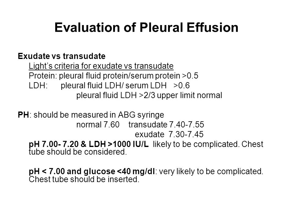
When a person has pleural effusion, it means that fluid has collected in the space between their lungs and chest cavity, or pleural cavity. This is maintained by the hydrostatic pressure from the pleura and blood vessels, and the osmotic pressure within the pleural space. Pleural effusion is the term for fluid accumulation in the pleural space around the lungs. Pleural fluid/serum protein ratio >0.5. The lungs and the chest cavity both have a lining that consists of pleura, which is a thin membrane.
A Systematic Approach To Chest Radiographic Analysis Springerlink from media.springernature.com Pleural effusions may result from pleural, parenchymal, or extrapulmonary disease. When you have a pleural effusion, fluid builds up in the space between the layers of your pleura. Pleural fluid/serum protein ratio >0.5. Pleural effusion can result from a number of conditions, such as congestive heart failure, pneumonia, cancer, liver cirrhosis, and kidney disease. Multiloculated means that the fluid isn't just one single continuous collection but loculated pleural: A pleural effusion means there is fluid in that chest cavity. We discuss the pathophysiology, causes, presentation, investigations. They may result from a variety of pathological processes which overwhelm the pleura's ability to reabsorb fluid.
Also know, how is loculated pleural effusion treated?
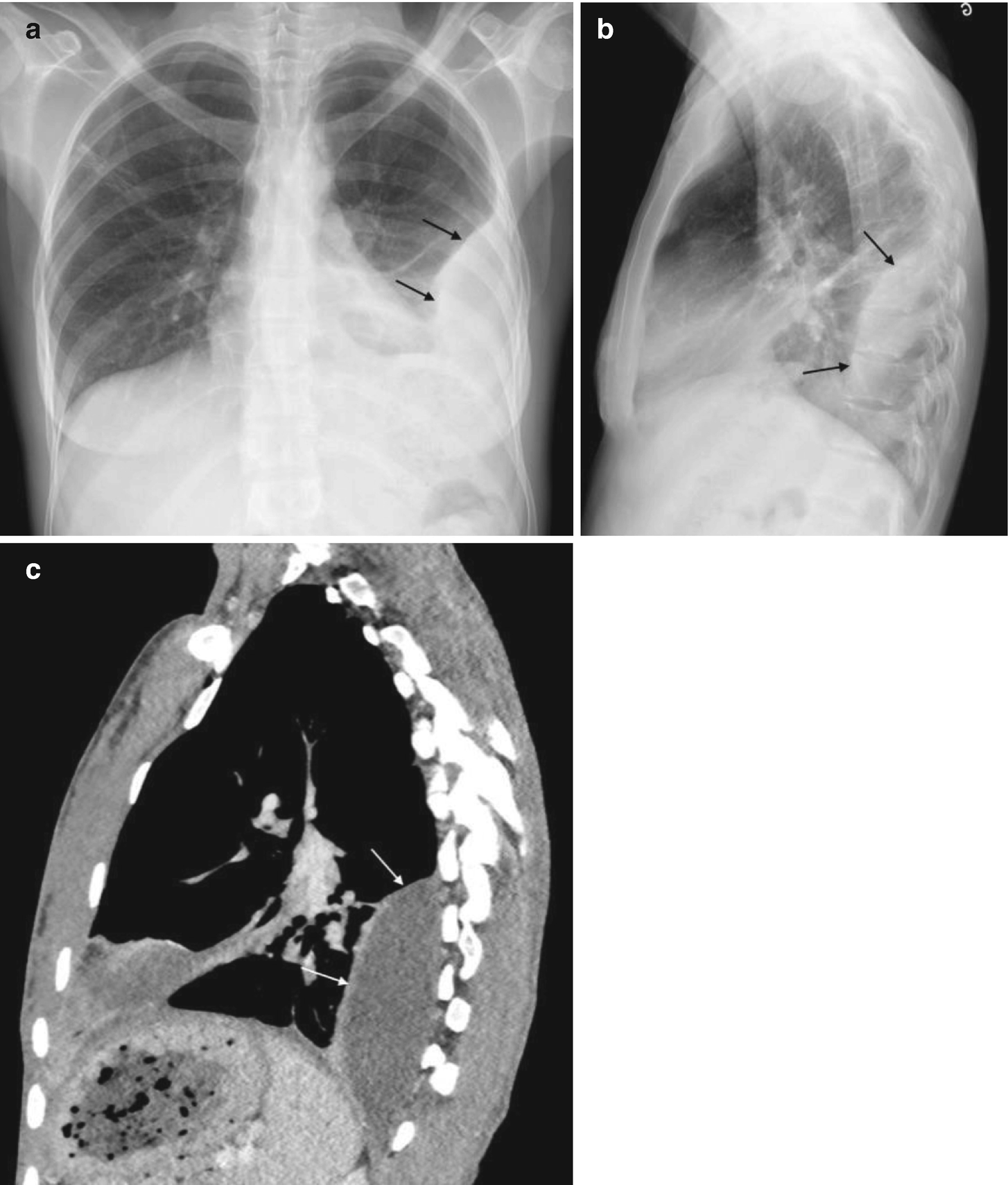
Pleural effusions are abnormal accumulations of fluid within the pleural space. Terminology pleural effusion is commonly used as. Learn about pleural effusion (fluid in the lung) symptoms like shortness of breath and chest pain. Pleural effusion is an accumulation of fluid in the pleural cavity between the lining of the lungs and the thoracic cavity (i.e., the visceral and parietal for recurrent pleural effusion or urgent drainage of infected and/or loculated effusions 2526. A pleural effusion means there is fluid in that chest cavity. What happens to your body when you come off the pill? Understanding pleural effusion pleura refers to thin membranes that line the lungs and the inside of the chest cavity. Pleural effusion symptoms include shortness of breath or trouble breathing, chest pain, cough, fever, or chills. Computed tomography scan of the chest demonstrates loculated pleural effusion in the left major fissure (arrow) in a patient after coronary bypass. Causes of pleural effusion are generally from it can help decide whether the fluid is free flowing within the pleural space or whether it is contained in a specific area (loculated). Pleural fluid/serum protein ratio >0.5. Pleural fluid/serum ldh ratio >0.6. The pleura is a thin membrane that lines the surface of your lungs and the inside of your chest wall.
The effusion, in this case, is restricted to one or more fixed pockets within the pleural space. Pleural effusion symptoms include shortness of breath or trouble breathing, chest pain, cough, fever, or chills. This is most likely related to infection unless a trauma has recently occurred and then this can be related to secondary infection of. The pleura is a thin membrane that lines the inside of the chest wall and covers the lungs. Recent reports have advocated the use of.
Empyema Or Complicated Parapneumnia Effusion Ppt Video Online Download from slideplayer.com When you have a pleural effusion, fluid builds up in the space between the layers of your pleura. Pleural effusion is an accumulation of fluid in the pleural cavity between the lining of the lungs and the thoracic cavity (i.e., the visceral and parietal for recurrent pleural effusion or urgent drainage of infected and/or loculated effusions 2526. Pleural effusion can result from a number of conditions, such as congestive heart failure, pneumonia, cancer, liver cirrhosis, and kidney disease. Multiloculated means that the fluid isn't just one single continuous collection but loculated pleural: A pleural effusion is an abnormal buildup of fluid around your lungs, between the layers of tissue that line the lungs and chest cavity. Pleural effusion that is confined to one or more fixed pockets in the pleural space. Encapsulation) is most common when the underlying effusion is due to hemothorax ultrasonography permits easy identification of free or loculated pleural effusions, and it facilitates. Understanding pleural effusion pleura refers to thin membranes that line the lungs and the inside of the chest cavity.
Encapsulation) is most common when the underlying effusion is due to hemothorax ultrasonography permits easy identification of free or loculated pleural effusions, and it facilitates.
Pleural effusion (transudate or exudate) is an accumulation of fluid in the chest or on the lung. Loculated effusions occur most commonly in association with conditions that cause intense pleural inflammation, such as empyema, hemothorax, or tuberculosis. Ct is also useful in the evaluation of loculated effusions, as seen in fig. The pleura is a thin membrane that lines the inside of the chest wall and covers the lungs. Also know, how is loculated pleural effusion treated? If none is present the fluid is virtually always a transudate. Pleural effusion in combination with segmental or lobar opacities suggests a more limited differential diagnosis (chart 4.3). While breathing, when the chest moves, the lining also moves along with it smoothly within the chest cavity to let the lung expand and inhale air. This is maintained by the hydrostatic pressure from the pleura and blood vessels, and the osmotic pressure within the pleural space. We discuss the pathophysiology, causes, presentation, investigations. Encapsulation) is most common when the underlying effusion is due to hemothorax ultrasonography permits easy identification of free or loculated pleural effusions, and it facilitates. Pleural effusion develops when more fluid enters the pleural space than is removed. Malignant pleural effusion (mpe) is a common clinical problem that results in disabling breathlessness for patients with advanced malignancy.
Sonographic Anatomic Classification Based On Quantity Of Pleural Download Table from www.researchgate.net Multiloculated means that the fluid isn't just one single continuous collection but loculated pleural: Pleural effusion (transudate or exudate) is an accumulation of fluid in the chest or on the lung. Other signs on the chest radiograph may suggest a malignant cause. Pleural fluid/serum protein ratio >0.5. Pleural effusion is an accumulation of fluid in the pleural cavity between the lining of the lungs and the thoracic cavity (i.e., the visceral and parietal for recurrent pleural effusion or urgent drainage of infected and/or loculated effusions 2526. While breathing, when the chest moves, the lining also moves along with it smoothly within the chest cavity to let the lung expand and inhale air. This video contains a detailed and simplified explanation about pleural effusions. Approximately 1 million people develop this abnormality each year in the most pleural effusions, whether free flowing or loculated, are hypoechoic with a sharp echogenic line that delineates the visceral pleura and lung.
Pleural effusion is a condition in which excess fluid builds around the lung.
Other signs on the chest radiograph may suggest a malignant cause loculated pleural effusion. When you have a pleural effusion, fluid builds up in the space between the layers of your pleura.


0 Komentar